
Successful implantation of the main cochlear implant under the microscope in otolaryngology surgery
Introduction: In the context of precision medicine, the application of ENT surgical microscope has brought revolutionary progress to the high-precision and difficult surgery of cochlear implantation. It not only significantly improves the success rate and safety of surgery, but also marks a new stage of visualization and minimally invasive otomicrosurgery.
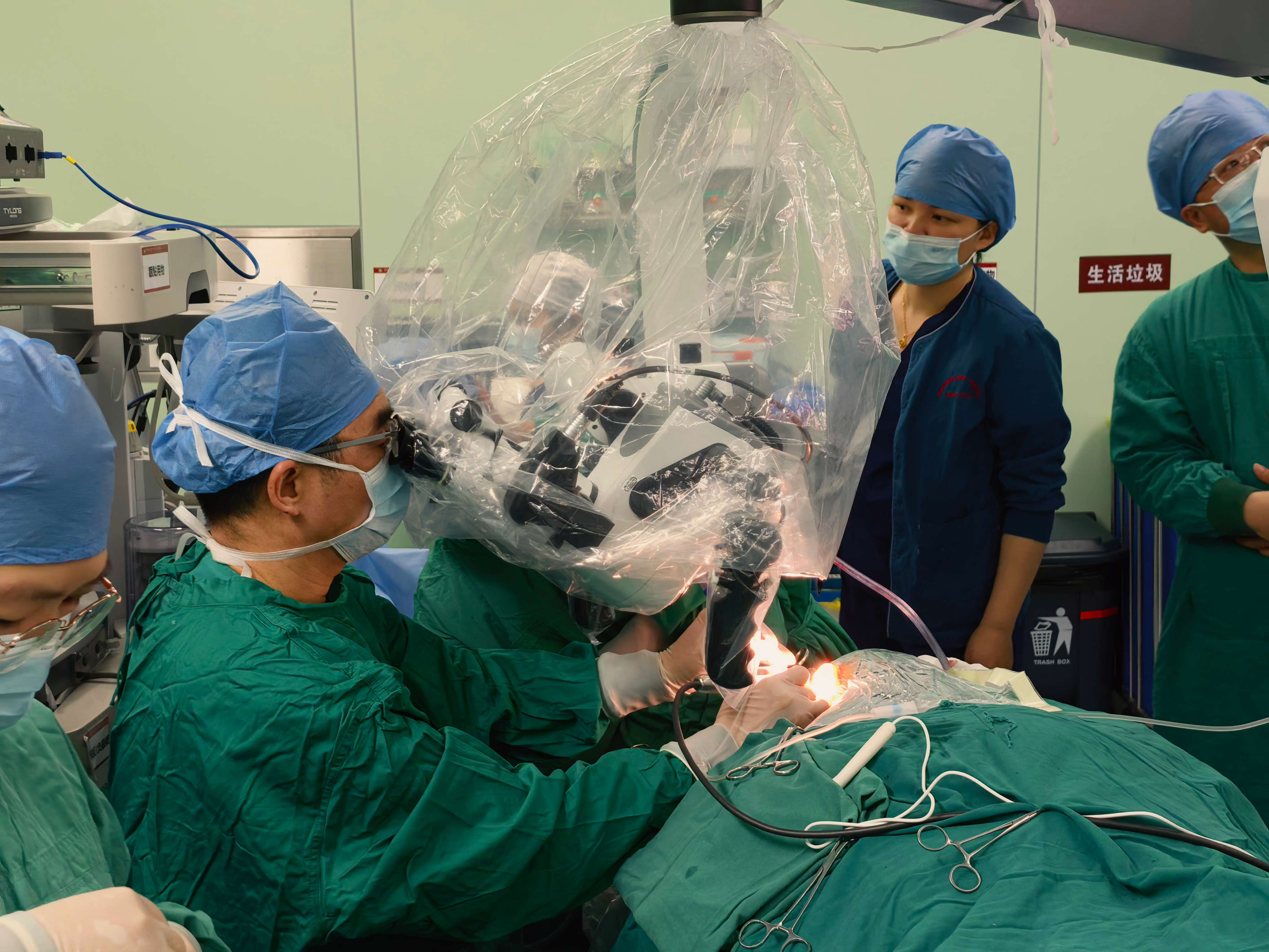
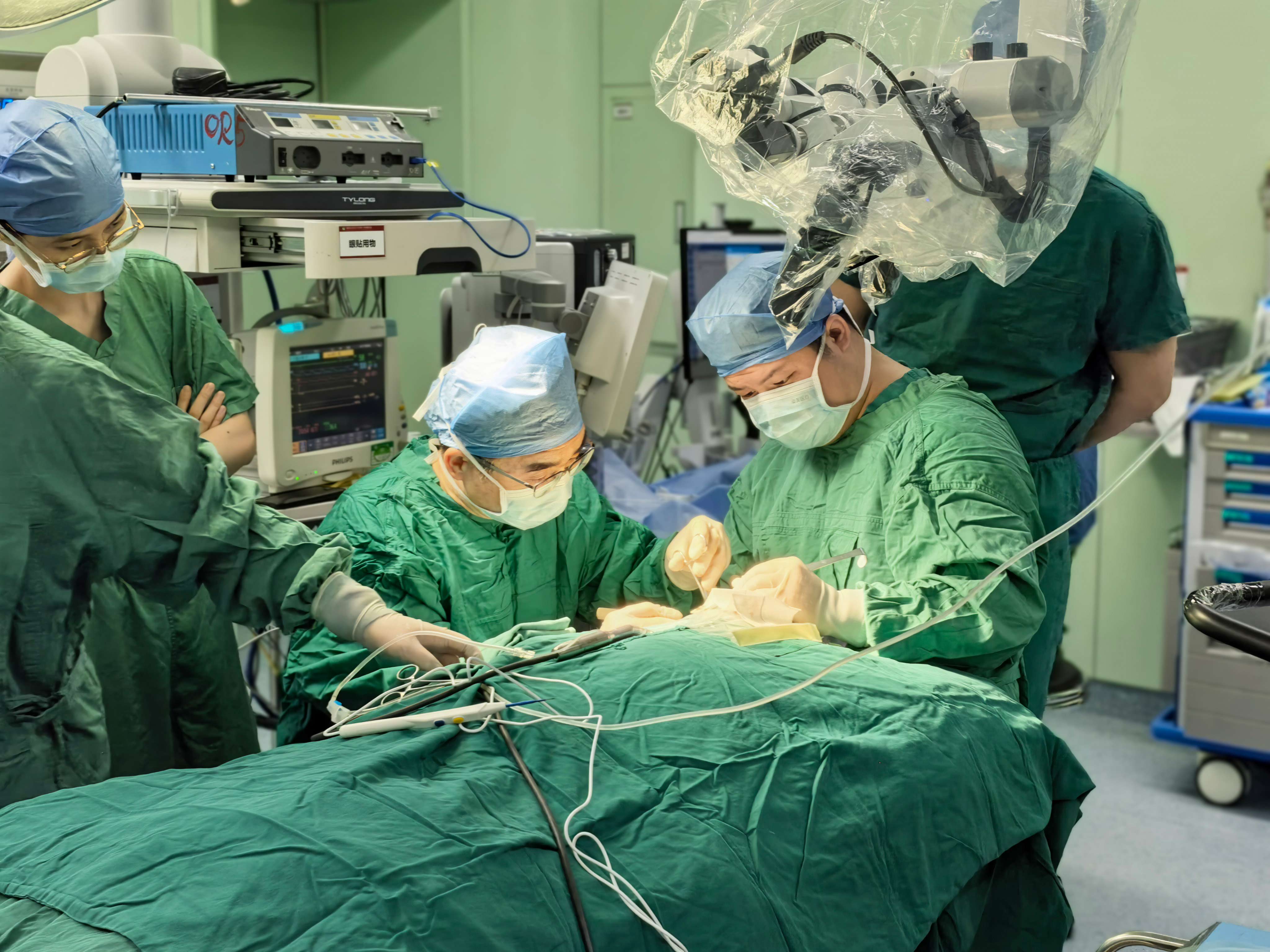
1 The 'smart eye' on the operating table: the core role of microscopes in cochlear implant surgery
Cochlear implantation is an effective method for treating severe to extremely severe sensorineural hearing loss. The surgery requires opening a small channel in the temporal bone behind the ear, accurately implanting the electrode array into the cochlear drum step, and avoiding damage to the facial nerve, fine structures of the inner ear, etc. This process imposes almost stringent requirements on the illumination, magnification, and fine manipulation of the surgical field.
Even with the help of headlights and magnifying glasses, the field of view and resolution of traditional surgical methods are still limited. And modern otolaryngology specialized surgical microscopes, with their excellent optical performance, have become the "third eye" of the chief surgeon. In surgery, it plays an irreplaceable and crucial role:
Providing a stereoscopic magnified field of view: The ENT microscope can provide continuous optical zoom from 6x to 40x, presenting the microstructure of the middle and inner ear (such as the round window membrane, stapes, facial nerve recess, etc.) clearly and stereoscopically in front of the surgeon, which is the visual basis for achieving precise implantation.
Realize coaxial cold light illumination: Its fiber optic light source system can directly project high-intensity cold light into deep and narrow surgical fields (such as facial crypts), with almost no shadows, ensuring a bright and uniform operating area, greatly improving tissue recognition.
Supporting fine manual operation: Under a stable and enlarged field of view, doctors can confidently use microscopic instruments to perform precise steps such as bone grinding, opening facial crypts, cutting open circular window membranes, and gently implanting electrodes, minimizing trauma to surrounding tissues.
2 Clear, stable, and minimally invasive: the three core advantages brought by surgical microscopes
The application of otolaryngology surgical microscope in cochlear implantation has the advantages of comprehensive and significant:
A leap in precision and safety: The high-definition magnified field of view enables doctors to clearly distinguish important anatomical landmarks and variations, effectively avoiding damage to the facial nerve, chordal drum nerve, and internal ear membrane structures, and minimizing the risk of surgical complications such as facial paralysis, taste disorders, and cerebrospinal fluid leakage. The implantation of electrodes is also more precise and smooth, which is beneficial for protecting residual nerve fibers in the cochlea and laying the foundation for better auditory effects after surgery.
True minimally invasive surgery: Under the surgical microscope, surgery can develop towards a more minimally invasive direction. For example, smaller skin incisions and more precise bone window opening (round window pathway or cochlear fenestration) can be achieved, reducing damage to the patient's physiological structure, resulting in less postoperative pain, faster recovery, smaller scars, and better cosmetic effects.
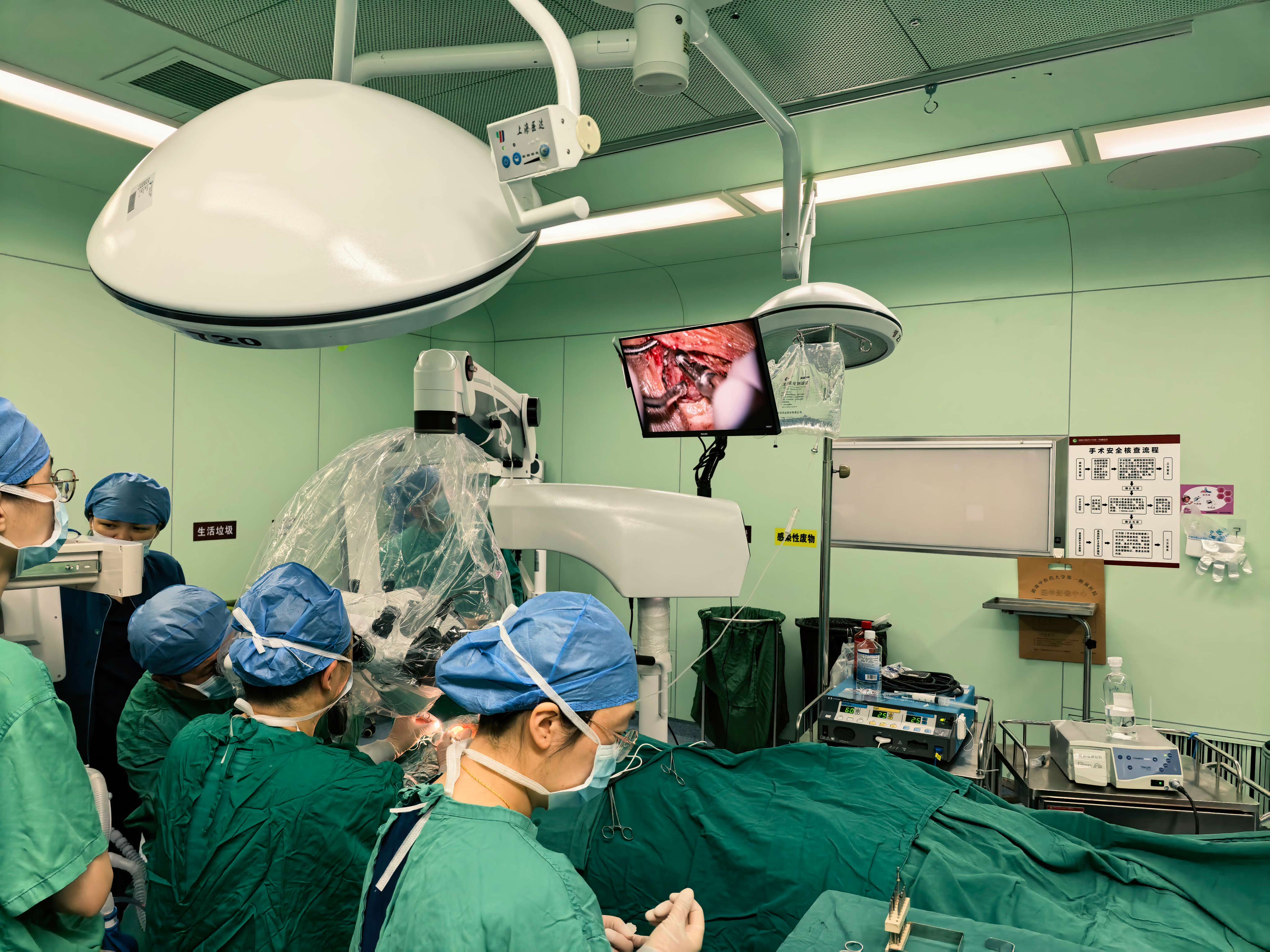
Improving surgical efficiency and teaching value: A stable field of view reduces visual fatigue for surgeons, making long-term surgical operations easier. At the same time, the video capture system of the microscope can record high-definition surgical images in real time, which not only facilitates intraoperative team collaboration, but also provides valuable materials for young doctors' observation and learning, postoperative review, and academic exchange.
3 From Macro to Micro: The Development Path of Medical Surgery Led by Surgical Microscopy
The success of cochlear implant surgery is a microcosm of the successful application of surgical microscopes in the field of otoneurosurgery, reflecting the profound development of modern surgical techniques
The paradigm shift from "big knife" to "minimally invasive intervention": Surgical microscopes are one of the core tools driving this transformation. It enables surgeons to go beyond the physiological limits of the human eye, perform fine operations in the microscopic world, and elevate surgical goals from "excising lesions" to "precise functional reconstruction".
Multi technology integration platform: Modern surgical microscopes are no longer just optical devices. It integrates digital cameras, fluorescence contrast imaging (such as ICG for observing blood vessels), optical coherence tomography (OCT) interfaces, and other functions, and is linked with neural monitoring and surgical navigation systems, becoming the "information center" in surgical procedures, promoting the development of intelligent and diversified diagnosis and treatment integration in surgery.
Empowering the popularization of difficult surgeries: With the assistance of microscopes, many surgeries that were previously impossible or extremely risky (such as cochlear implantation, stapes surgery, acoustic neuroma resection, etc.) have been standardized and regulated, allowing more patients to receive high-quality treatment in a wider range of medical institutions, which has significant social significance.
Conclusion
The successful application of otolaryngology surgical microscope in cochlear implantation surgery is a model of modern technology empowering precision medicine. It is like a silent 'super assistant', using light and shadow as a pen to depict the medical miracle of regaining a new 'voice' in every inch. With the further integration of optics, digital technology, and artificial intelligence, future surgical microscopes will become smarter and more powerful, continuing to lead ear surgery and even minimally invasive surgery towards greater precision, safety, and humanity, bringing light and hope to more patients.
Post time: Apr-02-2026


