
The development of optical imaging in video based surgical microscopes
In the field of medicine, surgery is undoubtedly the core means of treating the vast majority of diseases, especially playing a crucial role in the early treatment of cancer. The key to the success of a surgeon's surgery lies in the clear visualization of the pathological section after dissection. Surgical microscopes have been widely used in medical surgery due to their strong sense of three dimensionality, high definition, and high resolution. However, the anatomical structure of the pathological part is intricate and complex, and most of them are adjacent to important organ tissues. The millimeter to micrometer structures have far exceeded the range that can be observed by the human eye. In addition, the vascular tissue in the human body is narrow and crowded, and the lighting is insufficient. Any small deviation may cause harm to the patient, affect the surgical effect, and even endanger life. Therefore, researching and developing Operating microscopes with sufficient magnification and clear visual images is a topic that researchers continue to explore in depth.
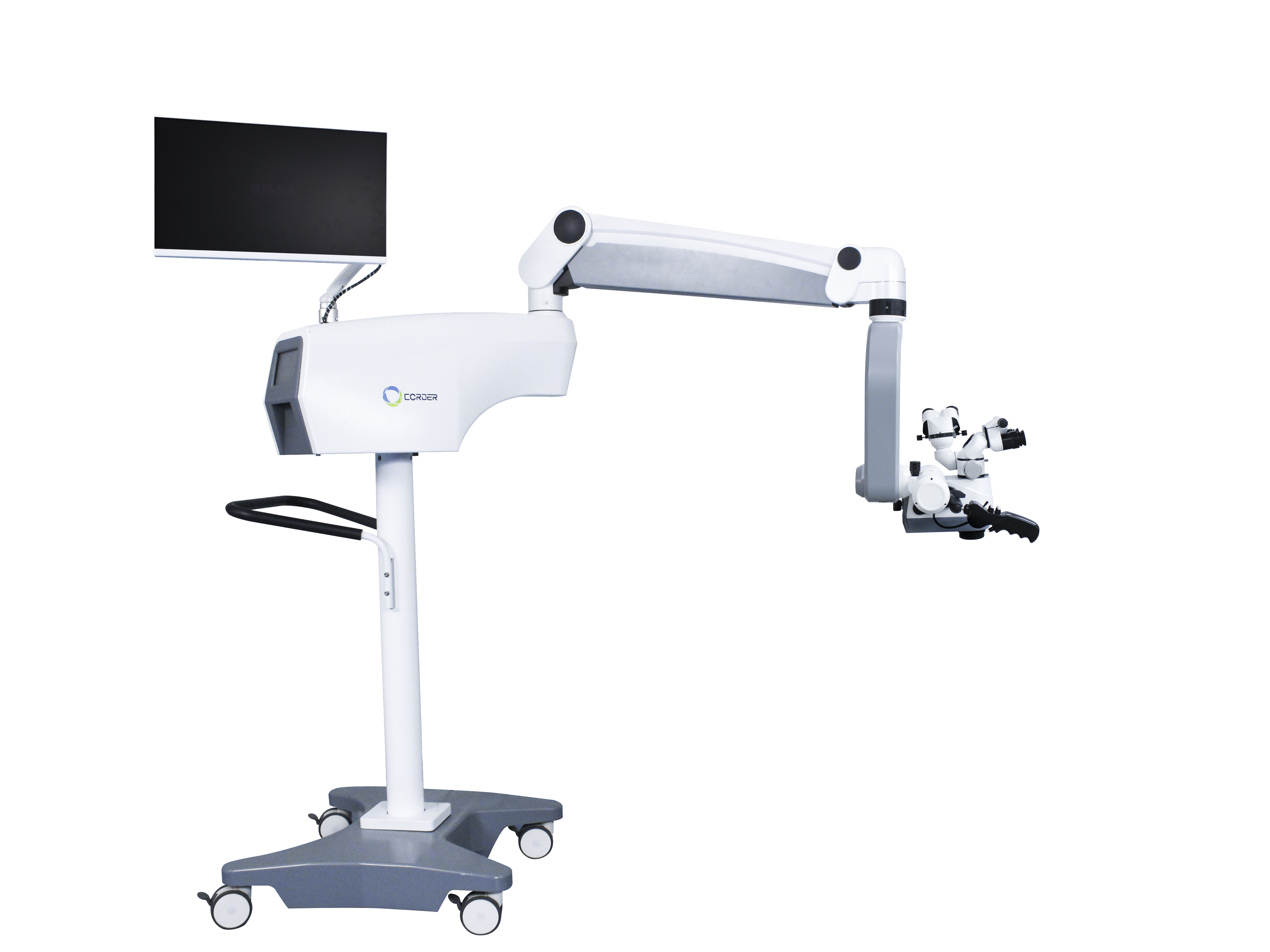
Currently, digital technologies such as image and video, information transmission, and photographic recording are entering the field of microsurgery with new advantages. These technologies are not only profoundly influencing human lifestyles, but also gradually integrating into the field of microsurgery. High definition displays, cameras, etc. can effectively meet the current requirements for surgical accuracy. Video systems with CCD, CMOS and other image sensors as receiving surfaces have gradually been applied to surgical microscopes. Video surgical microscopes are highly flexible and convenient for doctors to operate. The introduction of advanced technologies such as navigation system, 3D display, high-definition image quality, augmented reality (AR), etc., which enable multi person view sharing during the surgical process, further assists doctors in better performing intraoperative operations.
Microscope optical imaging is the main determinant of microscope imaging quality. The optical imaging of video surgical microscopes has unique design features, using advanced optical components and imaging technologies such as high-resolution, high contrast CMOS or CCD sensors, as well as key technologies such as optical zoom and optical compensation. These technologies effectively improve the imaging clarity and quality of microscopes, providing good visual assurance for surgical operations. Moreover, by combining optical imaging technology with digital processing, real-time dynamic imaging and 3D reconstruction have been achieved, providing surgeons with a more intuitive visual experience. In order to further improve the optical imaging quality of video surgical microscopes, researchers are constantly exploring new optical imaging methods, such as fluorescence imaging, polarization imaging, multispectral imaging, etc., to enhance the imaging resolution and depth of microscopes; Utilizing artificial intelligence technology for post-processing of optical imaging data to enhance image clarity and contrast.
In early surgical procedures, binocular microscopes were mainly used as auxiliary tools. A binocular microscope is an instrument that uses prisms and lenses to achieve stereoscopic vision. It can provide depth perception and stereoscopic vision that monocular microscopes do not have. In the mid-20th century, von Zehender pioneered the application of binocular magnifying glasses in medical ophthalmic examinations. Subsequently, Zeiss introduced a binocular magnifying glass with a working distance of 25 cm, laying the foundation for the development of modern microsurgery. In terms of optical imaging of binocular surgical microscopes, the working distance of early binocular microscopes was 75 mm. With the development and innovation of medical instruments, the first surgical microscope OPMI1 was introduced, and the working distance can reach 405 mm. The magnification is also constantly increasing, and the magnification options are constantly increasing. With the continuous advancement of binocular microscopes, their advantages such as vivid stereoscopic effect, high clarity, and long working distance have made binocular surgical microscopes widely used in various departments. However, the limitation of its large size and small depth cannot be ignored, and medical staff need to frequently calibrate and focus during surgery, which increases the difficulty of operation. In addition, surgeons who focus on visual instrument observation and operation for a long time not only increase their physical burden, but also do not comply with ergonomic principles. Doctors need to maintain a fixed posture to perform surgical examinations on patients, and manual adjustments are also required, which to some extent increases the difficulty of surgical operations.
After the 1990s, camera systems and image sensors began to gradually integrate into surgical practice, demonstrating significant application potential. In 1991, Berci innovatively developed a video system for visualizing surgical areas, with an adjustable working distance range of 150-500 mm and observable object diameters ranging from 15-25 mm, while maintaining a depth of field between 10-20 mm. Although the high maintenance costs of lenses and cameras at the time limited the widespread application of this technology in many hospitals, researchers continued to pursue technological innovation and began to develop more advanced video based surgical microscopes. Compared to binocular surgical microscopes, which require a long period of time to maintain this unchanged working mode, it can easily lead to physical and mental fatigue. The video type surgical microscope projects the magnified image onto the monitor, avoiding prolonged poor posture of the surgeon. Video based surgical microscopes liberate doctors from a single posture, allowing them to operate on anatomical sites through high-definition screens.
In recent years, with the rapid advancement of artificial intelligence technology, surgical microscopes have gradually become intelligent, and video based surgical microscopes have become mainstream products in the market. The current video based surgical microscope combines computer vision and deep learning technologies to achieve automated image recognition, segmentation, and analysis. During the surgical process, intelligent video based surgical microscopes can assist doctors in quickly locating diseased tissues and improving surgical accuracy.
In the development process from binocular microscopes to video based surgical microscopes, it is not difficult to find that the requirements for accuracy, efficiency, and safety in surgery are increasing day by day. Currently, the demand for optical imaging of surgical microscopes is not limited to magnifying pathological parts, but is increasingly diversified and efficient. In clinical medicine, surgical microscopes are widely used in neurological and spinal surgeries through fluorescence modules integrated with augmented reality. AR navigation system can facilitate complex spinal keyhole surgery, and fluorescent agents can guide doctors to completely remove brain tumors. In addition, researchers have successfully achieved automatic detection of vocal cord polyps and leukoplakia using a hyperspectral surgical microscope combined with image classification algorithms. Video surgical microscopes have been widely used in various surgical fields such as thyroidectomy, retinal surgery, and lymphatic surgery by combining with fluorescence imaging, multispectral imaging, and intelligent image processing technologies.
Compared to binocular surgical microscopes, video microscopes can provide multi-user video sharing, high-definition surgical images, and are more ergonomic, reducing doctor fatigue. The development of optical imaging, digitization, and intelligence has greatly improved the performance of surgical microscope optical systems, and real-time dynamic imaging, augmented reality, and other technologies have greatly expanded the functions and modules of video based surgical microscopes.
The optical imaging of future video based surgical microscopes will be more precise, efficient, and intelligent, providing doctors with more comprehensive, detailed, and three-dimensional patient information to better guide surgical operations. Meanwhile, with the continuous advancement of technology and the expansion of application fields, this system will also be applied and developed in more fields.
Post time: Nov-07-2025


