
A Simplified Guide to the Use of Neurosurgical Microscopes
Neurosurgical microscopes are essential tools used in neurosurgery to provide high-quality magnification and visualization during delicate procedures. In this guide, we will explain the key components, proper setup, and basic operation of a neurosurgical microscope. The aim is to provide a simplified understanding so that both medical professionals and interested readers can grasp its usage.
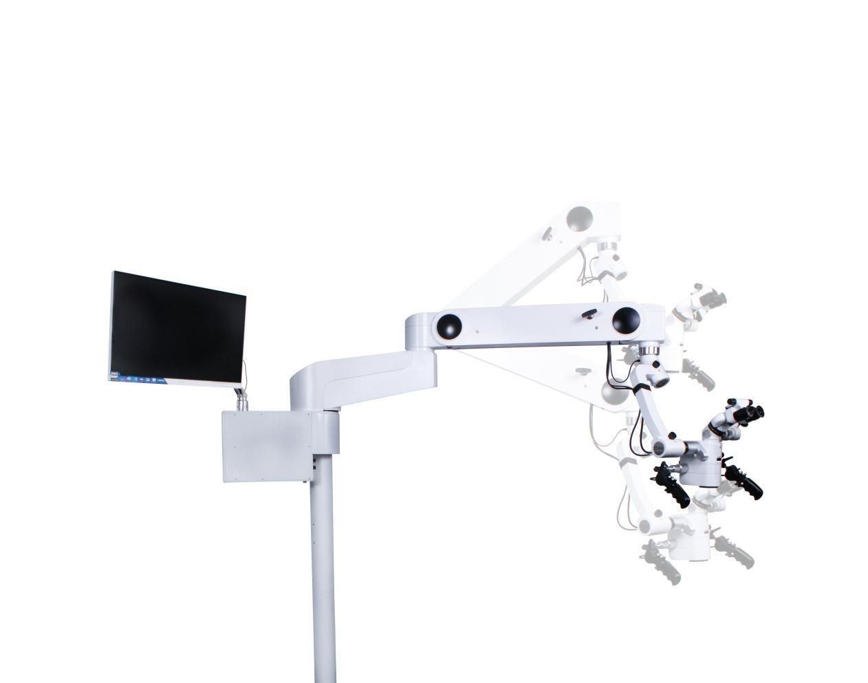
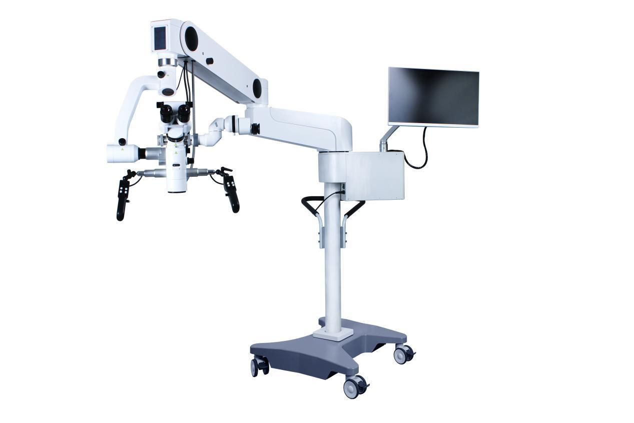
Overview of the Neurosurgical Microscope The neurosurgical microscope comprises several main components. Firstly, there is the optical system, which includes the objective lens and oculars (eyepieces) that magnify the surgical field. The microscope's stand or mount supports the optical system and allows for stable positioning. Next, the illumination system provides bright light to enhance visibility, usually through a fiberoptic cable or LED lighting. Finally, various accessories such as filters, zoom controls, and focusing mechanisms are available to optimize the microscope's functionality.
Proper Setup of the Neurosurgical Microscope Before starting the procedure, it is crucial to set up the microscope correctly. Begin by attaching the microscope to a sturdy base or tripod. Align the objective lens with the center of the microscope's field of view. Adjust the height and tilt of the microscope to ensure a comfortable working position. Connect the illumination system, ensuring a uniform and focused light beam onto the surgical field. Finally, calibrate the microscope's working distance and magnification levels according to the specific surgical requirements.
Basic Operation and Usage To begin using the neurosurgical microscope, position the patient correctly on the operating table and align the microscope's optical system with the surgical site. Using the focusing mechanisms, obtain a sharp focus on the region of interest. Adjust the magnification level to achieve the desired level of detail. Throughout the procedure, it is essential to maintain a sterile field by using sterile drapes and covers on the microscope. Additionally, be cautious when moving or adjusting the position of the microscope to avoid any unintended disturbance to the surgical field.
Advanced Features and Functions Neurosurgical microscopes offer various advanced features to enhance precision and accuracy during surgeries. Many models provide features such as digital imaging capabilities, allowing surgeons to capture and record high-resolution images or videos for documentation or educational purposes. Some microscopes also offer filters to enhance specific tissue visualization, such as fluorescence filters. Understandably, each microscope model may have its own unique set of features, and it is advisable to consult the manufacturer's manual to fully utilize these advanced functions.
Precautions and Maintenance Like any sophisticated medical equipment, neurosurgical microscopes require regular maintenance and care. It is essential to clean and disinfect the microscope after each use, following the manufacturer's guidelines to avoid damage to the delicate optical components. Regular servicing by qualified professionals is also recommended to ensure the microscope's optimal performance. Additionally, avoid exposing the microscope to excessive heat, moisture, or direct sunlight, as these can impair its functionality.
In conclusion, the neurosurgical microscope is an indispensable tool in modern neurosurgery, providing enhanced visualization and magnification during complex procedures. Understanding the basic setup, operation, and maintenance of the microscope is vital for efficient and effective usage. By following these guidelines, medical professionals can leverage the capabilities of the neurosurgical microscope to improve patient outcomes and safety.
Post time: Aug-03-2023


