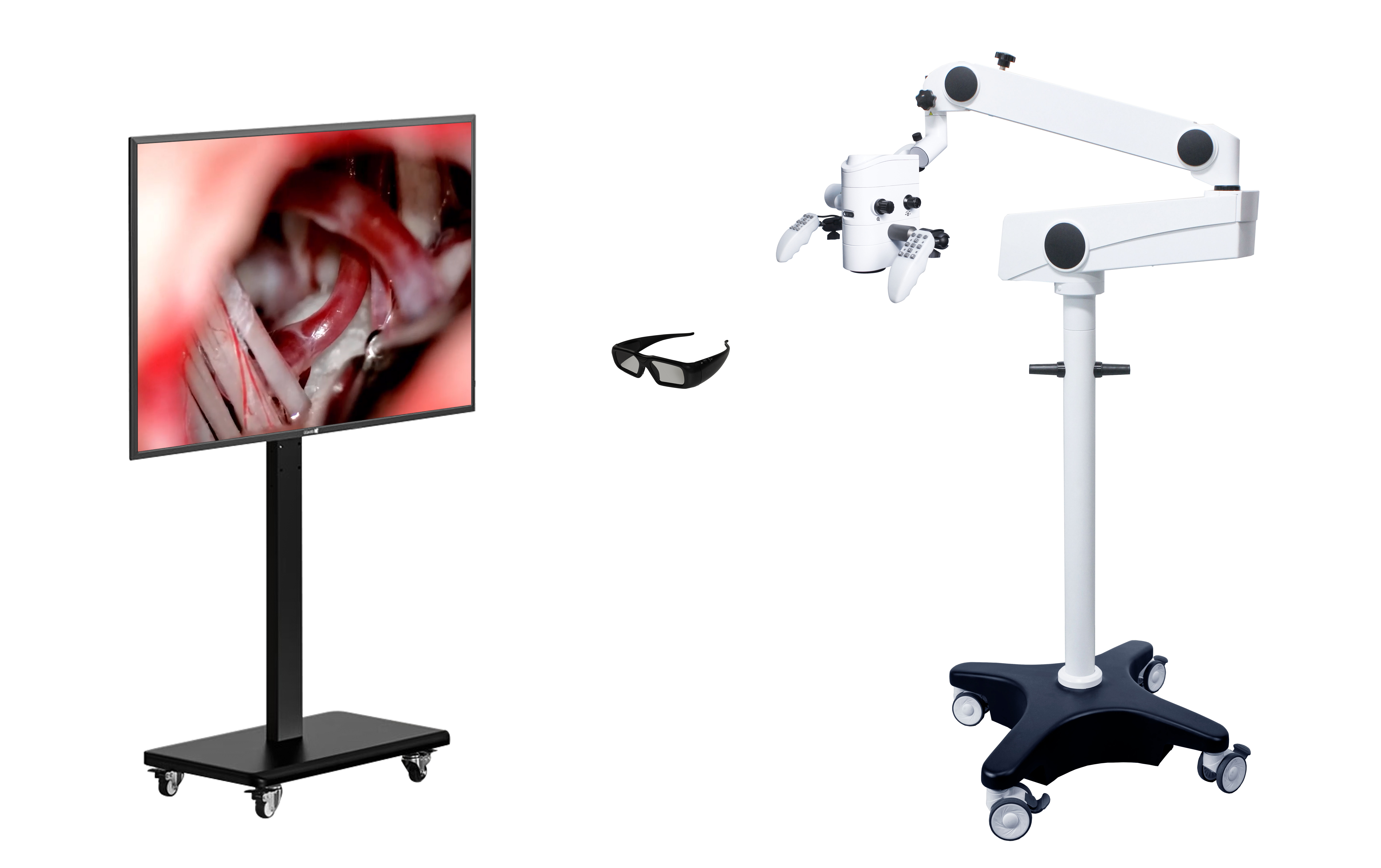
Progress of application of exoscopes in neurosurgical procedures
The application of surgical microscopes and neuroendoscopes has notably enhanced the efficacy of neurosurgical procedures, Nevertheless, owing to some inherent characteristics of the equipment themselves, they stilhave certain constraints in clinical applications. ln light of the deficiencies of operating microscopes and neuroendoscopes, coupled with the progressions in digital imaging, Wifi network connectivity, screen technology and optical technology, the exoscope system has come into being as a bridge between surgical microscopes and neuroendoscopes. The exoscope possesses superior image quaity and surgical visual field, better ergonomic posture, teaching efficacy as well as more efficient surgical team engagement, and its application eficacy is similar to that of strical microscopes. At present, the literature mainly reports the disparities between exoscopes and surgical microscopes in technicaequipment aspects such as depth of field, visual field, focal length and operation, lacking a summary and analysis of the specific applcation and surgical outcomes of exoscopes in neurosurgery, Hence, we summarize the application exoscopes in neurosurgery in recent years, analyze their advantages and limitations in clinical practice, and offer references for cinical utilization.
The History and Development of exoscopes
Surgical microscopes have excellent deep illumination, high-resolution surgical field of view, and stereoscopic imaging effects, which can help surgeons observe the deep neural and vascular tissue structure of the surgical field more clearly and improve the accuracy of microscopic operations. However, the depth of field of the surgical microscope is shallow and the field of view is narrow, especially at high magnification. The surgeon needs to repeatedly focus and adjust the angle of the target area, which has a significant impact on the surgical rhythm; On the other hand, the surgeon needs to observe and operate through a microscope eyepiece, requiring the surgeon to maintain a fixed posture for a long time, which can easily lead to fatigue. In the past few decades, minimally invasive surgery has rapidly developed, and neuroendoscopic systems have been widely used in neurosurgery due to their high-quality images, better clinical outcomes, and higher patient satisfaction. However, due to the narrow channel of the endoscopic approach and the presence of important neurovascular structures near the channel, coupled with the characteristics of cranial surgery such as the inability to expand or shrink the cranial cavity, neuroendoscopy is mainly used for skull base surgery and ventricular surgery via nasal and oral approaches.
Given the shortcomings of surgical microscopes and neuroendoscopes, coupled with advances in digital imaging, WiFi network connectivity, screen technology, and optical technology, the external mirror system has emerged as a bridge between surgical microscopes and neuroendoscopes. Similar to neuroendoscopy, the external mirror system usually consists of a farsightedness mirror, a light source, a high-definition camera, a display screen, and a bracket. The main structure that distinguishes external mirrors from neuroendoscopy is a farsightedness mirror with a diameter of about 10 mm and a length of about 140 mm. Its lens is at a 0 ° or 90 ° angle to the long axis of the mirror body, with a focal length range of 250-750 mm and a depth of field of 35-100 mm. The long focal length and deep depth of field are the key advantages of external mirror systems over neuroendoscopy.
The advancement of software and hardware technology has promoted the development of exterior mirrors, especially the emergence of 3D exterior mirrors, as well as the latest 3D 4K ultra high definition exterior mirrors. The exterior mirror system is constantly updated every year. In terms of software, the external mirror system can visualize the surgical area by integrating preoperative magnetic resonance diffusion tensor imaging, intraoperative navigation, and other information, thereby helping doctors perform precise and safe surgeries. In terms of hardware, the external mirror can integrate 5-aminolevulinic acid and indocyanine filters for angiography, pneumatic arm, adjustable operating handle, multi screen output, longer focusing distance and larger magnification, thereby achieving better image effects and operating experience.
Comparison between exoscope and surgical microscopes
The external mirror system combines the external features of neuroendoscopy with the image quality of surgical microscopes, complementing each other's strengths and weaknesses, and filling the gaps between surgical microscopes and neuroendoscopy. External mirrors have the characteristics of deep depth of field and wide field of view (surgical field diameter of 50-150 mm, depth of field of 35-100 mm), providing extremely convenient conditions for deep surgical operations under high magnification; On the other hand, the focal length of the external mirror can reach 250-750mm, providing a longer working distance and facilitating surgical operations [7]. Regarding the visualization of external mirrors, Ricciardi et al. found through comparison between external mirrors and surgical microscopes that external mirrors have comparable image quality, optical power, and magnification effects to microscopes. The external mirror can also quickly switch from a microscopic perspective to a macroscopic perspective, but when the surgical channel is "narrow at the top and wide at the bottom" or obstructed by other tissue structures, the field of view under the microscope is usually limited. The advantage of the external mirror system is that it can perform surgery in a more ergonomic posture, reducing the time spent viewing the surgical field through the microscope eyepiece, thereby reducing the doctor's surgical fatigue. The external mirror system provides the same quality 3D surgical images to all surgical participants during the surgical process. The microscope allows up to two people to operate through the eyepiece, while the external mirror can share the same image in real time, allowing multiple surgeons to perform surgical operations simultaneously and improving surgical efficiency by sharing information with all personnel. At the same time, the external mirror system does not interfere with the mutual communication of the surgical team, allowing all surgical personnel to participate in the surgical process.
exoscope in neurosurgery surgery
Gonen et al. reported 56 cases of glioma endoscopic surgery, of which only 1 case had complications (bleeding in the surgical area) during the perioperative period, with an incidence rate of only 1.8%. Rotermund et al. reported 239 cases of transnasal transsphenoidal surgery for pituitary adenomas, and the endoscopic surgery did not result in serious complications; Meanwhile, there was no significant difference in surgical time, complications, or resection range between endoscopic surgery and microscopic surgery. Chen et al. reported that 81 cases of tumors were surgically removed through the retrosigmoid sinus approach. In terms of surgical time, degree of tumor resection, postoperative neurological function, hearing, etc., endoscopic surgery was similar to microscopic surgery. Comparing the advantages and disadvantages of two surgical techniques, the external mirror is similar or superior to the microscope in terms of video image quality, surgical field of view, operation, ergonomics, and surgical team participation, while the depth perception is rated as similar or inferior to the microscope.
exoscope in Neurosurgery Teaching
One of the main advantages of external mirrors is that they allow all surgical personnel to share the same quality 3D surgical images, allowing all surgical personnel to participate more in the surgical process, communicate and transmit surgical information, facilitate teaching and guidance of surgical operations, increase teaching participation, and improve the effectiveness of teaching. Research has found that compared to surgical microscopes, the learning curve of external mirrors is relatively shorter. In laboratory training for suturing, when students and resident physicians receive training on both the endoscope and microscope, most students find it easier to operate with the endoscope. In the teaching of craniocervical malformation surgery, all students observed three-dimensional anatomical structures through 3D glasses, enhancing their understanding of craniocervical malformation anatomy, improving their enthusiasm for surgical operations, and shortening the training period.
Outlook
Although the external mirror system has made significant progress in application compared to microscopes and neuroendoscopes, it also has its limitations. The biggest drawback of early 2D external view mirrors was the lack of stereoscopic vision in magnifying deep structures, which affected surgical operations and surgeon judgment. The new 3D external mirror has improved the problem of lack of stereoscopic vision, but in rare cases, wearing polarized glasses for a long time can cause discomfort such as headache and nausea for the surgeon, which is the focus of technical improvement in the next step. In addition, in endoscopic cranial surgery, it is sometimes necessary to switch to a microscope during the operation because some tumors require fluorescence guided visual resection, or the depth of surgical field illumination is insufficient. In addition, in endoscopic cranial surgery, it is sometimes necessary to switch to a microscope during the operation because some tumors require fluorescence guided visual resection, or the depth of surgical field illumination is insufficient. Due to the high cost of equipment with special filters, fluorescence endoscopes have not yet been widely used for tumor resection. During surgery, the assistant stands in the opposite position to the chief surgeon, and sometimes sees a rotating display image. Using two or more 3D displays, the surgical image information is processed by software and displayed on the assistant screen in a flipped 180 ° form, which can effectively solve the problem of image rotation and enable the assistant to participate in the surgical process more conveniently.
In summary, the increasing use of endoscopic systems in neurosurgery represents the beginning of a new era of intraoperative visualization in neurosurgery. Compared with surgical microscopes, external mirrors have better image quality and surgical field of view, better ergonomic posture during surgery, better teaching effectiveness, and more efficient surgical team participation, with similar surgical outcomes. Therefore, for most common cranial and spinal surgeries, an endoscope is a safe and effective new option. With the advancement and development of technology, more intraoperative visualization tools can assist in surgical operations to achieve lower surgical complications and better prognosis.

Post time: Sep-08-2025


