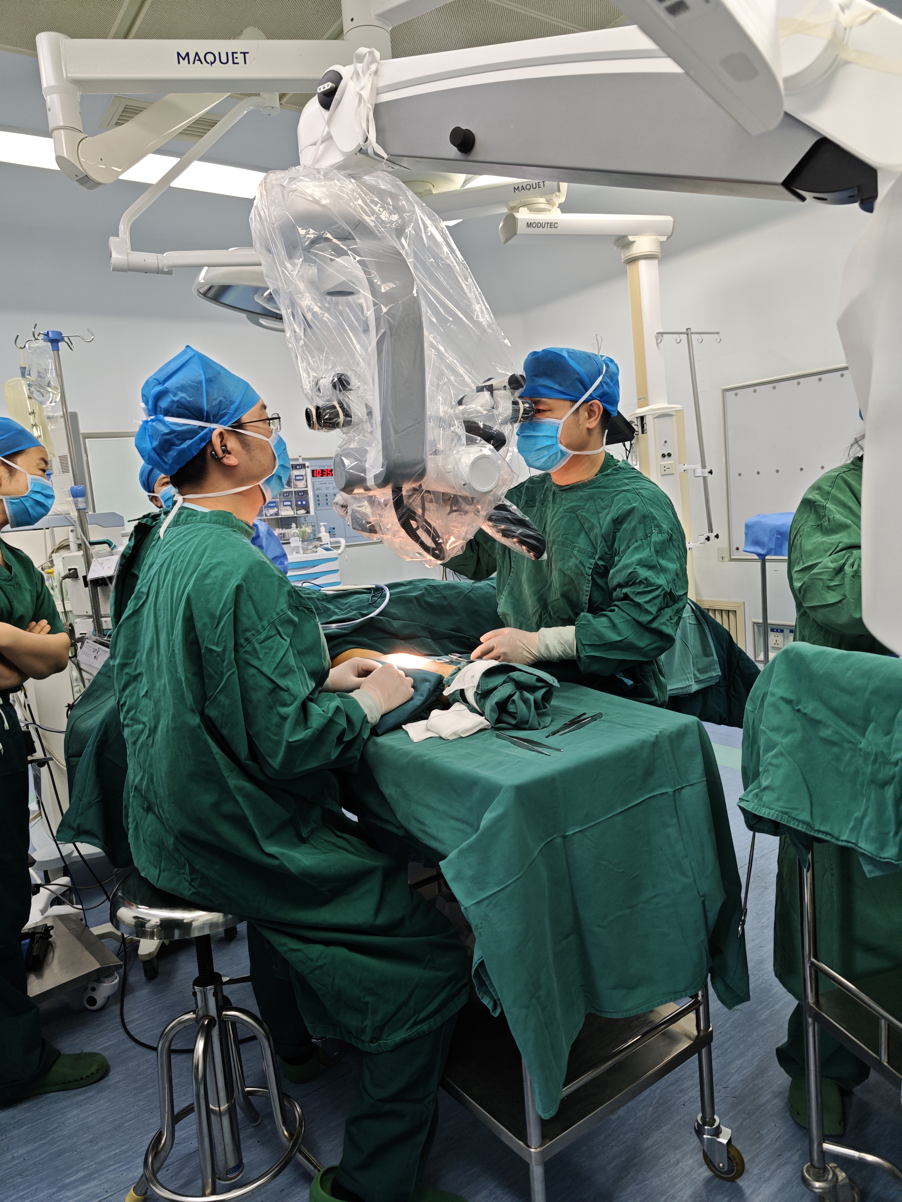
Technological Advancements and Clinical Applications of Ultra-High-Definition Surgical Microscopes
Surgical microscopes play an extremely important role in modern medical fields, especially in high-precision fields such as neurosurgery, ophthalmology, otolaryngology, and minimally invasive surgery, where they have become indispensable basic equipment. With high magnification capabilities, Operating microscopes provide a detailed view, allowing surgeons to observe details that are invisible to the naked eye, such as nerve fibers, blood vessels, and tissue layers, thereby helping doctors avoid damaging healthy tissue during surgery. Especially in neurosurgery, the high magnification of the microscope allows for precise localization of tumors or diseased tissues, ensuring clear resection margins and avoiding damage to critical nerves, thereby improving the quality of patients' postoperative recovery.
Traditional surgical microscopes are typically equipped with display systems of standard resolution, capable of providing sufficient visual information to support complex surgical needs. However, with the rapid development of medical technology, especially breakthroughs in the field of visual technology, the imaging quality of surgical microscopes has gradually become an important factor in improving surgical precision. Compared to traditional surgical microscopes, ultra-high-definition microscopes can present more details. By introducing display and imaging systems with resolutions of 4K, 8K, or even higher, ultra-high-definition surgical microscopes enable surgeons to more accurately identify and manipulate tiny lesions and anatomical structures, greatly enhancing the precision and safety of surgery. With the continuous integration of image processing technology, artificial intelligence, and virtual reality, ultra-high-definition surgical microscopes not only improve imaging quality but also provide more intelligent support for surgery, driving surgical procedures towards higher precision and lower risk.
Clinical application of ultra-high-definition microscope
With the continuous innovation of imaging technology, ultra-high-definition microscopes are gradually playing a pivotal role in clinical applications, thanks to their extremely high resolution, excellent imaging quality, and real-time dynamic observation capabilities.
Ophthalmology
Ophthalmic surgery requires precise operation, which imposes high technical standards on ophthalmic surgical microscopes. For instance, in femtosecond laser corneal incision, the surgical microscope can provide high magnification to observe the anterior chamber, central incision of the eyeball, and check the position of the incision. In ophthalmic surgery, illumination is crucial. The microscope not only provides optimal visual effects with lower light intensity but also produces a special red light reflection, which aids in the entire cataract surgery process. Furthermore, optical coherence tomography (OCT) is widely used in ophthalmic surgery for subsurface visualization. It can provide cross-sectional images, overcoming the limitation of the microscope itself, which cannot see fine tissues due to frontal observation. For example, Kapeller et al. used a 4K-3D display and a tablet computer to automatically stereoscopically display the effect diagram of Microscope-integrated OCT (miOCT) (4D-miOCT). Based on user subjective feedback, quantitative performance evaluation, and various quantitative measurements, they demonstrated the feasibility of using a 4K-3D display as a substitute for 4D-miOCT on a white light microscope. Additionally, in Lata et al.'s study, by collecting cases of 16 patients with congenital glaucoma accompanied by bull's eye, they used a microscope with miOCT function to observe the surgical process in real time. By evaluating key data such as preoperative parameters, surgical details, postoperative complications, final visual acuity, and corneal thickness, they ultimately showed that miOCT can help doctors identify tissue structures, optimize operations, and reduce the risk of complications during surgery. However, despite OCT gradually becoming a powerful auxiliary tool in vitreoretinal surgery, especially in complex cases and novel surgeries (such as gene therapy), some doctors question whether it can truly improve clinical efficiency due to its high cost and long learning curve.
Otolaryngology
Otorhinolaryngology surgery is another surgical field that utilizes surgical microscopes. Due to the presence of deep cavities and delicate structures in the facial features, magnification and illumination are crucial for surgical outcomes. Although endoscopes can sometimes provide a better view of narrow surgical areas, ultra-high-definition surgical microscopes offer depth perception, allowing for magnification of narrow anatomical regions such as the cochlea and sinuses, assisting doctors in treating conditions like otitis media and nasal polyps. For instance, Dundar et al. compared the effects of microscope and endoscope methods for stapes surgery in the treatment of otosclerosis, collecting data from 84 patients diagnosed with otosclerosis who underwent surgery between 2010 and 2020. Using the change in air-bone conduction difference before and after surgery as the measurement indicator, the final results showed that although both methods had similar effects on hearing improvement, surgical microscopes were easier to operate and had a shorter learning curve. Similarly, in a prospective study conducted by Ashfaq et al., the research team performed microscope-assisted parotidectomy on 70 patients with parotid gland tumors between 2020 and 2023, focusing on evaluating the role of microscopes in facial nerve identification and protection. The results indicated that microscopes had significant advantages in improving surgical field clarity, accurately identifying the main trunk and branches of the facial nerve, reducing nerve traction, and hemostasis, making them an important tool for enhancing facial nerve preservation rates. Furthermore, as surgeries become increasingly complex and precise, the integration of AR and various imaging modes with surgical microscopes enables surgeons to perform image-guided surgeries.
Neurosurgery
The application of ultra-high-definition surgical microscopes in neurosurgery has shifted from traditional optical observation to digitalization, augmented reality (AR), and intelligent assistance. For instance, Draxinger et al. utilized a microscope combined with a self-developed MHz-OCT system, providing high-resolution three-dimensional images through a 1.6 MHz scanning frequency, successfully assisting surgeons in distinguishing between tumors and healthy tissues in real time and enhancing surgical precision. Hafez et al. compared the performance of traditional microscopes and the ultra-high-definition microsurgical imaging system (Exoscope) in experimental cerebrovascular bypass surgery, finding that although the microscope had shorter suture times (P<0.001), the Exoscope performed better in terms of suture distribution (P=0.001). Additionally, the Exoscope provided a more comfortable surgical posture and shared vision, offering pedagogical advantages. Similarly, Calloni et al. compared the application of the Exoscope and traditional surgical microscopes in the training of neurosurgery residents. Sixteen residents performed repetitive structural recognition tasks on cranial models using both devices. The results showed that although there was no significant difference in overall operation time between the two, the Exoscope performed better in identifying deep structures and was perceived as more intuitive and comfortable by most participants, with the potential to become mainstream in the future. Evidently, ultra-high-definition surgical microscopes, equipped with 4K high-definition displays, can provide all participants with better-quality 3D surgical images, facilitating surgical communication, information transfer, and improving teaching efficiency.
Spinal surgery
Ultra-high-definition surgical microscopes play a pivotal role in the field of spinal surgery. By providing high-resolution three-dimensional imaging, they enable surgeons to observe the complex anatomical structure of the spine more clearly, including subtle parts such as nerves, blood vessels, and bone tissues, thereby enhancing the precision and safety of surgery. In terms of scoliosis correction, surgical microscopes can improve the clarity of surgical vision and fine manipulation ability, helping doctors accurately identify neural structures and diseased tissues within the narrow spinal canal, thus safely and effectively completing decompression and stabilization procedures.
Sun et al. compared the efficacy and safety of microscope-assisted anterior cervical surgery and traditional open surgery in the treatment of ossification of the posterior longitudinal ligament of the cervical spine. Sixty patients were divided into the microscope-assisted group (30 cases) and the traditional surgery group (30 cases). The results showed that the microscope-assisted group had superior intraoperative blood loss, hospital stay, and postoperative pain scores compared to the traditional surgery group, and the complication rate was lower in the microscope-assisted group. Similarly, in spinal fusion surgery, Singhatanadgige et al. compared the application effects of orthopedic surgical microscopes and surgical magnifying glasses in minimally invasive transforaminal lumbar fusion. The study included 100 patients and showed no significant differences between the two groups in postoperative pain relief, functional improvement, spinal canal enlargement, fusion rate, and complications, but the microscope provided a better field of view. In addition, microscopes combined with AR technology are widely used in spinal surgery. For example, Carl et al. established AR technology in 10 patients using the head-mounted display of a surgical microscope. The results showed that AR has great potential for application in spinal degenerative surgery, especially in complex anatomical situations and resident education.
Summary and Outlook
Compared to traditional surgical microscopes, ultra-high-definition surgical microscopes offer numerous advantages, including multiple magnification options, stable and bright illumination, precise optical systems, extended working distances, and ergonomic stable stands. Furthermore, their high-resolution visualization options, especially the integration with various imaging modes and AR technology, effectively support image-guided surgeries.
Despite the numerous advantages of surgical microscopes, they still face significant challenges. Due to their bulky size, ultra-high-definition surgical microscopes pose certain operational difficulties during transportation between operating rooms and intraoperative positioning, which may adversely affect the continuity and efficiency of surgical procedures. In recent years, the structural design of microscopes has been significantly optimized, with their optical carriers and binocular lens barrels supporting a wide range of tilt and rotational adjustments, greatly enhancing the operational flexibility of the equipment and facilitating the surgeon's observation and operation in a more natural and comfortable position. Furthermore, the continuous development of wearable display technology provides surgeons with more ergonomic visual support during microsurgical operations, helping to alleviate operational fatigue and improve surgical precision and the surgeon's sustained performance ability. However, due to the lack of a supporting structure, frequent refocusing is required, making the stability of wearable display technology inferior to that of conventional surgical microscopes. Another solution is the evolution of equipment structure towards miniaturization and modularization to adapt more flexibly to various surgical scenarios. However, volume reduction often involves precision machining processes and high-cost integrated optical components, making the actual manufacturing cost of the equipment expensive.
Another challenge of ultra-high-definition surgical microscopes is skin burns caused by high-power illumination. To provide bright visual effects, especially in the presence of multiple observers or cameras, the light source must emit strong light, which may burn the patient's tissue. It has been reported that ophthalmic surgical microscopes can also cause phototoxicity to the ocular surface and tear film, leading to decreased ocular cell function. Therefore, optimizing light management, adjusting the spot size and light intensity according to magnification and working distance, is particularly important for surgical microscopes. In the future, optical imaging may introduce panoramic imaging and three-dimensional reconstruction technologies to expand the field of view and accurately restore the three-dimensional layout of the surgical area. This will enable doctors to better understand the overall situation of the surgical area and avoid missing important information. However, panoramic imaging and three-dimensional reconstruction involve real-time acquisition, registration, and reconstruction of high-resolution images, generating huge amounts of data. This places extremely high demands on the efficiency of image processing algorithms, hardware computing power, and storage systems, especially during surgery where real-time performance is crucial, making this challenge even more prominent.
With the rapid development of technologies such as medical imaging, artificial intelligence, and computational optics, ultra-high-definition surgical microscopes have demonstrated great potential in enhancing surgical precision, safety, and operational experience. In the future, ultra-high-definition surgical microscopes may continue to develop in the following four directions: (1) In terms of equipment manufacturing, miniaturization and modularization should be achieved at lower costs, making large-scale clinical application possible; (2) Develop more advanced light management modes to address the issue of light damage caused by prolonged surgery; (3) Design intelligent auxiliary algorithms that are both precise and lightweight to meet the computational performance requirements of the equipment; (4) Deeply integrate AR and robotic surgical systems to provide platform support for remote collaboration, precise operation, and automated processes. In summary, ultra-high-definition surgical microscopes will evolve into a comprehensive surgical assistance system that integrates image enhancement, intelligent recognition, and interactive feedback, helping to build a digital ecosystem for future surgery.
This article provides an overview of the advancements in common key technologies of ultra-high-definition surgical microscopes, with a focus on their application and development in surgical procedures. With the enhancement of resolution, ultra-high-definition microscopes are playing a pivotal role in fields such as neurosurgery, ophthalmology, otolaryngology, and spinal surgery. Especially, the integration of intraoperative precision navigation technology in minimally invasive surgeries has elevated the precision and safety of these procedures. Looking ahead, as artificial intelligence and robotic technologies advance, ultra-high-definition microscopes will offer more efficient and intelligent surgical support, propelling the progression of minimally invasive surgeries and remote collaboration, thereby further elevating surgical safety and efficiency.

Post time: Sep-05-2025


