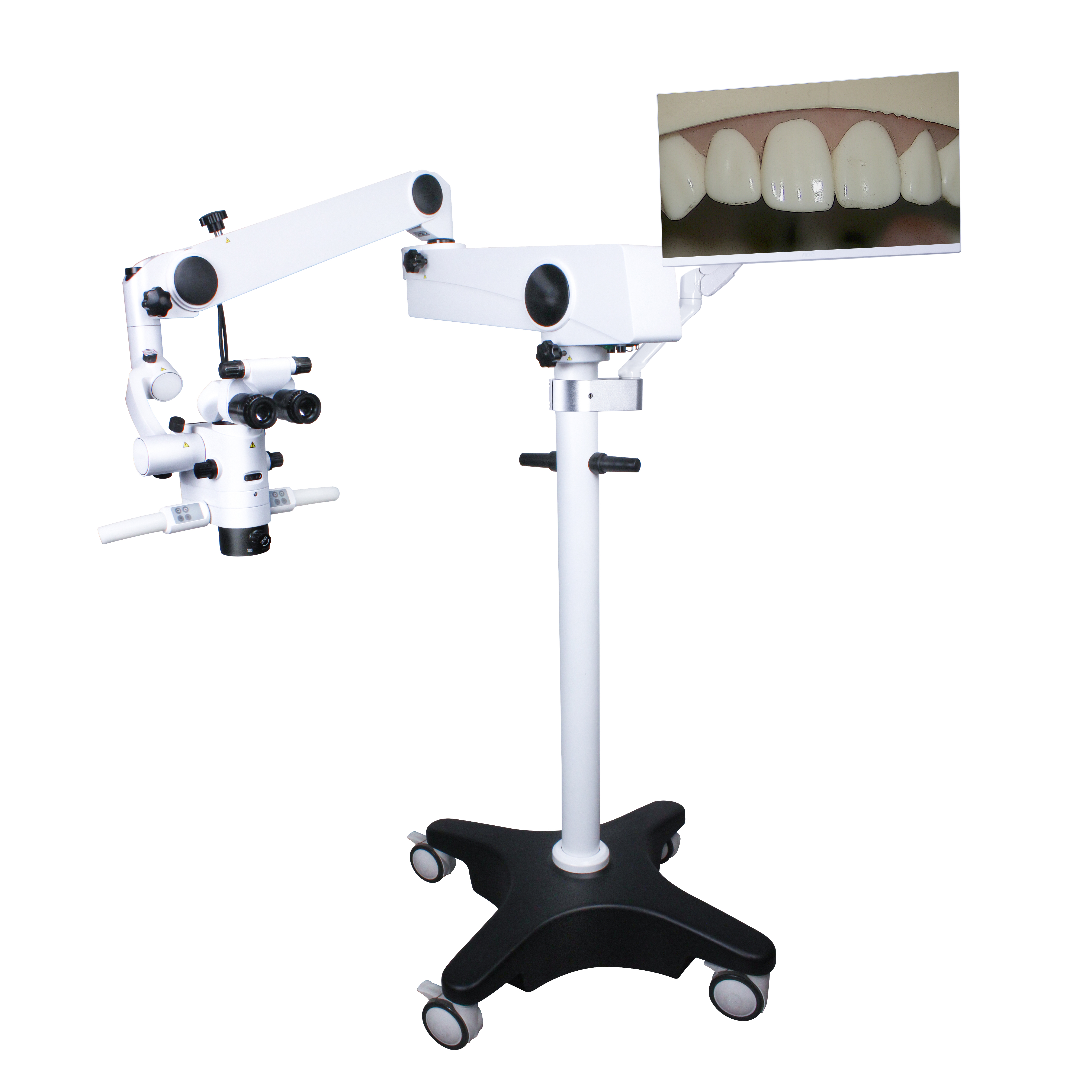
Revolution in Dental Pulp Treatment under Microscopic Perspective: Practical Experience and Insights from a Clinical Doctor
When I first started practicing, I relied on my sense of touch and experience to "blindly explore" in a narrow field of vision, and often regretfully declared tooth extraction due to the complexity of the root canal system that I could not directly see. It was not until the introduction of Dental Surgical Microscope that a new dimension of precise dental pulp treatment was truly opened up. This device is not simply an amplifier - its LED Microscope Light Source provides cold light source shadowless illumination that penetrates deep into the medullary cavity, while the High Resolution Microscope Camera projects the root canal isthmus, accessory sulcus, and even microcracks onto a high-definition screen, turning diagnosis from speculation to evidence. For example, a calcified lower molar that was deemed "unable to locate the root canal" by an exceptional hospital showed enamel color difference at the MB2 root canal opening under 25 times magnification. With the help of an ultrasound working tip, it was successfully cleared, avoiding the risk of lateral perforation caused by excessive cutting.
In Microscope Surgery, the operational logic is completely refactored. Traditional root canal re treatment relies on hand feel to remove broken instruments, which can easily cause displacement or perforation; Under the Microscope Operation, I used a micro file with the assistance of ultrasonic vibration to gradually unwind around the top of the broken needle, visualizing the entire process to ensure maximum preservation of dentin. For cracked teeth, the application of Microscopios Dental has further overturned the prognosis: shallow cracks that were previously easily missed by staining and probing can now be clearly presented in terms of direction and depth under the microscope. I have performed minimally invasive resin filling and full crown restoration on 58 cases of shallow hidden cracked teeth, with a success rate of 79.3%. Among them, 12 cases were promptly converted to root canal treatment due to early microscopic detection of cracks extending to the pulp floor, avoiding the risk of subsequent fracture.
The value of Microscope Surgery is particularly prominent in periapical surgery. A patient with recurrent periapical abscess underwent traditional surgery that required flap removal to expose a large area of the surgical site, while Microscopic Operation used a 4k Camera Microscope for real-time navigation under a local small flap window to accurately remove 3mm of the periapical abscess and prepare it backwards. The tightness of MTA backfilling was verified to be seamless at 400x magnification. The postoperative bone defect area was filled with artificial bone powder, and a one-year follow-up showed complete bone regeneration and normal dental function. The success rate of such cases can reach over 90%, far higher than the 60% -70% of traditional surgery, which confirms the revolutionary promotion of microscopy technology for the goal of "tooth preservation".
However, the advantages of the equipment need to be matched with the abilities of the surgeon. Systematic Dental Microscope Training is the core threshold - from position adjustment, pupillary distance calibration to multi-level zoom switching, overcoming dizziness and hand eye coordination barriers. I initially trained on the model for 20 hours before adapting to the depth control of Dental Microscope, but in actual practice, clearing calcified root canals requires a cumulative total of more than 50 operations to achieve a stable success rate. It is recommended that new scholars start with marrow opening and root canal positioning, gradually advancing to complex operations such as perforation repair.
Choosing an A Good Microscope requires comprehensive consideration. There are many Microscope Brands, but the core parameters need to be focused: the objective focal length is above 200mm to ensure the operating space, the zoom range is 3-30x to adapt to different surgical procedures, and the lifespan of the Microscope LED Light Source should exceed 1000 hours to prevent intraoperative attenuation. In Parts Of A Microscope, multi angle binoculars and electric focusing modules are essential, otherwise frequent manual adjustments will interrupt the treatment process. Which Microscope To Buy? It is recommended to prioritize evaluating optical performance over additional features. For example, although a certain brand's basic model does not have a built-in camera, it can still meet teaching needs when paired with an external 4K Camera Microscope; However, excessive pursuit of magnification may sacrifice the width of the field of view, which is not conducive to clinical efficiency. The price of Microeye Microscope is often affected by configuration, with mid-range models costing around 200000 to 400000 yuan, but 10% of the budget needs to be reserved for maintenance. Purchase through legitimate Microscope Retailers to ensure that warranty terms cover critical services such as optical path calibration. The after-sales response speed of Microscope Companies and Fabricantes De Microscopios Endodonticos also needs to be evaluated - if lens fouling or joint lock failure cannot be resolved within 48 hours, it will lead to delayed diagnosis and treatment.
Nowadays, dental operating microscope has become my "third eye" for daily diagnosis and treatment. It reconstructs the treatment standards: from the thoroughness of root canal cleaning to the tightness of the repair edge, the microscopic accuracy constantly refreshes the definition of 'success'. When my peers consulted Buy Microscope for advice, I emphasized that this is not only a device upgrade, but also a reshaping of clinical philosophy - only by infusing microscopic thinking into every operational detail can true minimally invasive treatment be achieved in the micrometer world.

Post time: Aug-15-2025


