Ophthalmoscope
Gonioscopy
Gonio Super m1-XGM1
With high magnification, the trabecular meshwork can be observed in detail.
The all-glass design provides exceptional clarity and durability.
Using angle examination and laser treatment, combined with the use of fundus laser, fundus photocoagulation.
|
Model |
Field |
Magnification |
Laser Spot Magnification |
Contact Surface Diameter |
|
XGM1 |
62° |
1.5X |
0.67X |
14.5mm |
Gonio Super m3-XGM3
Three lens, all optical glass, 60° lens provide a view of the iris angle
60° provides a retinal image from the equator to the ora serrata
76° mirror can see the middle peripheral/peripheral retina
|
Model |
Field |
Magnification |
Laser Spot Magnification |
Contact Surface Diameter |
|
XGM3 |
60°/66°/76° |
1.0X |
1.0X |
14.5mm |
Gonio Suspended Lens With Handle -XGSL
Combined with operating microscope, glaucoma surgery, all-optical glass lens body, excellent imaging quality. The suspendable mirror frame is convenient to adapt to the eye movement during the operation, stable imaging of the angle of the room, and ensures the quality of the angle surgery.
|
Model |
Magnification |
Handle Length |
Contact Lens Diameter |
Effective Caliber |
Positioning Diameter |
|
XGSL |
1.25X |
85mm |
9mm |
11mm |
14.5mm |
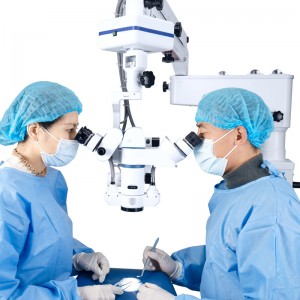
Eye Surgery Series
1.Use with microscope
Surgery 130WF NA -XO130WFN
Combined with surgical microscope, vitrectomy surgery, all-optical glass body, binocular aspheric surface, excellent imaging quality. Large viewing angle.
XO130WFN is Ethylene oxide disinfection.
|
Model |
Field |
Magnification |
contact lens diameter |
Lens barrel diameter |
|
XO130WFN |
112°-134° |
0.39x |
11.4mm |
21mm |
Surgery 130WF -XO130WF
Combined with surgical microscope, vitrectomy surgery, all-optical glass body, binocular aspheric surface, excellent imaging quality. Large viewing angle.
XO130WF sterilizes with high temperature and high pressure.
|
Model |
Field |
Magnification |
contact lens diameter |
Lens barrel diameter |
|
XO130WF |
112°-134° |
0.39x |
11.4mm |
21mm |
Special Purpose Series
Ldepth Vitreous - XIDV
Combined with ophthalmic laser, vitreous ablation laser surgery, all-optical glass mirror body, optical glass contact lens, excellent imaging quality. Treatment of fundus floaters.
| Model | Magnification | Laser Spot |
| XIDV | 1.18x | 0.85x |
Laser Iridectomy - XLIRIS
Combined with ophthalmic laser, iridotomy laser surgery, all-optical glass body, optical glass contact lens, excellent image quality. Wide-spectrum laser coating protective mirror.
| Model | Magnification | Laser Spot |
| XLIRIS | 1.67x | 0.6x |
Laser Capsulotomy - XLCAP
Combined with ophthalmic laser, capsulotomy laser surgery, all-optical glass body, optical glass contact lens, excellent imaging quality. Wide-spectrum laser coating protective mirror.
| Model | Magnification | Laser Spot |
| XLCAP | 1.6x | 0.63x |
Combined with fundus laser
XLP84-Laser posterior 84
Macular photocoagulation used, high magnification.
Ideal design for focused, gridded laser therapy.
Provides highly magnified images of the posterior pole of the eye and expands the field of view.
| Model | Field | Magnification | Laser Spot |
| XLP84 | 70°/84° | 1.05x | 0.95x |
XLC130-Laser Classic 130
For retinal detachments of the usual range.
High quality general diagnostic and laser therapy lenses.
Good PDT and PRP performance.
| Model | Field | Magnification | Laser Spot |
| XLC130 | 120°/144° | 0.55x | 1.82x |
XLM160-Laser mini 160
Smaller housing simplifies orbital manipulation.
Optical glass material, highest resolution imaging.
Good performance of PRP.
| Model | Field | Magnification | Laser Spot |
|
XLM160 |
156°/160° |
0.58x |
1.73X |
XLS165-Laser Super 165
Wide angle, good PRP performance.
Binocular aspheric surface, excellent image quality.
Curved mirror body for a comfortable grip.
| Model | Field | Magnification | Laser Spot |
| XLS165 | 160°/165° | 0.57x | 1.77x |
Fundus Examination
XSC90-Classic 90
Classic 90D optical glass material.
Suitable for small pupils, for general fundus examination.
Double aspherical lens enhance image, high-resolution stereoscopic image.
| Model | Field | Magnification | Laser Spot
Magnification |
Working distance |
|
XSC90 |
74°/ 89° | 0.76 | 1.32 | 7 mm |
XBC20-Classic 20
Classic 20D optical glass material
Use with binocular indirect ophthalmoscope
Fundus general examination
Double-aspherical lens
| Model | Field | Magnification | Laser Spot
Magnification |
Working distance |
| XBC20 | 46°-60° | 3.13 | 0.32 | 50mm |
XSS90-Super 90
Compared with Classic 90, the fundus area observed is larger.
Suitable for pan retinal examination.
Field of view increased to 116°.
| Model | Field | Magnification | Laser Spot
Magnification |
Working distance
|
| XSS90 | 95°/116° | 0.76 | 1.31 | 7 mm |
XSS78-Super 78
Use with slit lamp
Double-aspheric lens
excellent imaging quality
| Model | Field | Magnification | Laser Spot
Magnification |
Working distance
|
| XSS78 | 82°/98° | 1.05 | 0.95 | 10mm |
XSM90-Mater 90
Compared with Super90, the observed fundus area is larger.
The widest 124° and the widest field of view also maintain the same magnification.
Large imaging range and good uniformity.
| Model | Field | Magnification | Laser Spot
Magnification |
Working distance |
| XSM90 | 104°/125° | 0.72 | 1.39 | 4.5mm |
XSP90-Primary 90
Adopt new resin material, lighter and higher refractive index.
Super cost-effective.
Double-sided aspheric surface, eliminating spherical aberration and flare, excellent image quality.
| Model | Field | Magnification | Laser Spot
Magnification |
Working distance |
| XSP90 | 72°/ 86° | 0.82 | 1.22 | 7.5mm |
XSP78-Primary 78
Adopt new resin material, lighter and higher refractive index.
High magnification allows for excellent visualization of the optic disc and macula.
Fully corrected image curvature, astigmatism, aberration and coma
| Model | Field | Magnification | Laser Spot
Magnification |
Working distance |
| XSP78 | 82°/98° | 1.03 | 0.97 | 10mm |
Master Mag.
1.3x image magnification is the highest magnification of non-contact slit lamp lens
Double-sided aspheric surface, excellent image quality
High magnification, dedicated to the examination of fundus conditions in the macular area.
| Model | Field | Magnification | Laser Spot
Magnification |
Working distance |
| XSH50 | 66°/78° | 1.2 | 0.83 | 13mm |